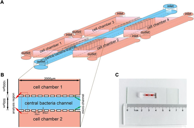
FIGURE 2.
Schematic depiction of a microfluidic device. (A) An illustration of the device for investigating bacterial chemotaxis mechanism. Microfluidic device constructed by central bacteria channel, four separate cell-culture chambers, and micro-channels. (B) The analysis region used to quantify the preferential accumulation of bacteria sited between two chambers. (C) An image of the integrated microfluidic device. Reproduced with permission from Song et al. (2018). Copyright 2018, Springer Nature.

