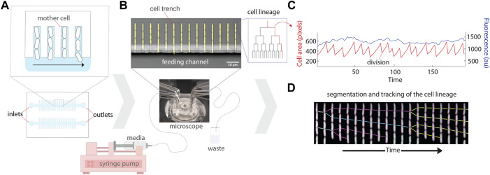
FIGURE 1.
Schematic of the experimental setup for the mother machine microfluidic device and data analysis. (A) Schematic representation of the platform which traps single bacterial cells in trenches that are perpendicular to a larger feeding channel. Daughter cells are flushed out of the trenches with flowing media, while mothers remain trapped at the end of the cell trench. (B) A micrograph of the mother machine, with YFP fluorescence showing the cells superimposed on a brightfield image of the device. Media is pumped through the inlet into the main feeding channel by a syringe pump, and then exits through the outlet into a waste beaker. (C) The lineages of growing cells in the trenches can then be followed under precisely controlled environmental conditions using time-lapse microscopy. (D) An example kymograph of a growing cell imaged in fluorescence, illustrating the segmentation and tracking of the lineage.

