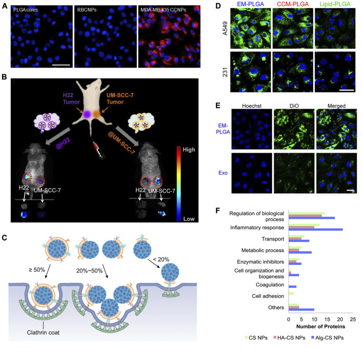
FIGURE 2.
Biological membranes and biomolecules of surface modification. (A) Fluorescent imaging of MDA-MB-435 cells incubated with PLGA cores, RBCM-coated PLGA NPs (RBCNPs), or MDA-MB-435 tumor CM-coated NPs (CCNPs). NPs were labeled with DiD (red) and nuclei were stained with DAPI (blue). Scale bar, 50 μm. Reprinted with permission from Fang et al. (2014). Copyright 2014 American Chemical Society. (B) Schematic illustration of the in vivo study with H22 and UM-SCC-7 dual-tumor-bearing mouse model. Fluorescence images and ex vivo images of tumors at 12 h post injection with H22 and UM-SCC-7 tumor CM-coated magnetic NPs. Reprinted with permission from Zhu et al. (2016). Copyright 2016 American Chemical Society. (C) Illustration of different cell internalization strategies by NPs with different CM coating percentages by Liu et al. (2021) licensed under CC BY 4.0. (D) Confocal fluorescence images of cellular uptake of A549 cell-derived EM-coated (EM-PLGA), A549 cancer CM-coated (CCM-PLGA), and lipid-coated (lipid-PLGA) PLGA NPs by A549 and MDA-MB-231 (231). The PLGA cores were labeled with DiO (green), and the cell nuclei were stained with Hoechst (blue). Scale bar, 20 μm. (E) Confocal fluorescence images showing higher cellular uptake of EM-PLGA NPs compared to exosomes (Exo) by A549 cells. The membranes of EM-PLGA NPs and Exo were labeled with DiO (green). The cell nuclei were stained with Hochest (blue). Scale bar, 40 μm; Reprinted with permission from (C. Liu et al. (2019)). Copyright 2019 American Chemical Society. (F) Comparison of functions of protein corona of bare chitosan NPs (CS NPs), HA-coated CS NPs (HA-CS NPs), and alginate-coated CS NPs (Alg-CS NPs) by (Almalik et al., 2017) licensed under CC BY 4.0.

