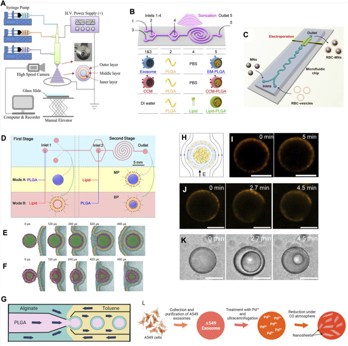
FIGURE 4.
Surface modification techniques of micro- and nano-scale biomaterials. (A) Schematic diagram of a tri-axial electrospray system to produce multilayer core-shell particles. Reprinted with permission from Yao et al. (2020). Copyright 2020 American Chemical Society. (B) Schematic representation of the microfluidics sonication method for synthesis of the A549 exosome membrane (EM)-, cancer cell membrane (CCM)-, and lipid-coated PLGA NPs through the combined effects of sonication and microfluidics. The microfluidics device consists of one straight channel and one spiral channel connected with four inlets (inlets 1–4) and one outlet (outlet 5). The device is immersed in an ultrasonic bath, and the generated EM-PLGA NPs, CCM-PLGA NPs, and lipid-PLGA NPs are collected from outlet 5. Reprinted with permission from (C Liu et al., 2019). Copyright 2019 American Chemical Society. (C) Microfluidics electroporation-facilitated synthesis of RBCM-coated magnetic NPs (RBC-MNs). Reprinted with permission from Rao et al. (2017). Copyright 2017 American Chemical Society. (D) Schematic representation of the two-stage microfluidics chip to produce monolayer (MP) and bilayer (BP) lipid shell for PLGA NPs. Reprinted with permission from Zhang et al. (2015). Copyright 2015 American Chemical Society. Use of molecular dynamics (MD) simulation to demonstrate the influence of rigidity in cellular uptake of (E) MP and (F) BP. Reprinted with permission from Sun et al. (2015). Copyright 2014 WILEY-VCH Verlag GmbH & Co. KGaA, Weinheim. (G) Schematic diagram showing fabrication of PLGA–alginate core–shell microspheres using a multi-capillary microfluidics chip (Wu et al., 2013). (H) Illustration and (I) confocal fluorescence images of fatty acid-coated coacervate droplets excited at 10 V/cm showing membrane (BODIPY 558/568 C12—orange) slipped at the direction of green arrows. (J) BODIPY 558/568 C12 (orange) labeled fatty acid-coated coacervate droplets at 20 V/cm and (K) simultaneous repetitive cycles of vacuolization at that voltage. Scale bar, 10 μm. Reprinted with permission from Jing et al. (2019). Copyright 2019 American Chemical Society. (L) Illustration of preparation of Pd-nanosheets encapsulated exosomes from A549 cells (Sancho-Albero et al., 2019a).

