Figure 6.
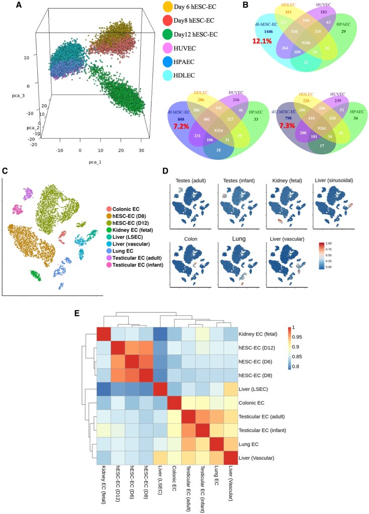
Transcriptomic comparison of human embryonic stem cell-derived endothelial cells to mature endothelial cells. (A) Three-dimensional principal component analysis plot of human embryonic stem cell-derived endothelial cells from d6, d8, and d12 datasets alongside mature endothelial cell: human dermal lymphatic endothelial cells, human umbilical vein endothelial cells, and human pulmonary artery endothelial cells. (B) Venn diagrams of number of genes expressed in d6, d8, and d12 human embryonic stem cell-derived endothelial cells and the overlap of these genes with each type of mature endothelial endothelial cell. Percentage of uniquely expressed human embryonic stem cell-derived endothelial cell genes shown in red. (C) tSNE plot showing clustering of endothelial cell extracted from 10X datasets of human organs. (D) Signature scores for each endothelial cell type shown in C. (E) Heatmap with hierarchical clustering comparing transcriptional status of each cell type by Spearman’s rank correlation coefficient.

