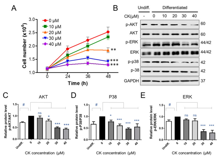
Figure 6.
The effect of CK on signaling molecules involved in mitotic regulation. (A) Two days after confluence, 3T3-L1 cells were incubated with DMI medium, with and without various concentrations of CK, for indicated times. The number of cells at the indicated points (0, 24, 36, and 48 h) after DMI-induced differentiation was counted using a Luna cell counting slide. The experiment was repeated independently three times. **: p < 0.01, ***: p < 0.001 compared to 0 µM CK group. (B) Protein levels of p-AKT, AKT, p-ERK, ERK, p-P38, and p38 during 48 h of differentiation were analyzed by immunoblot assay using specific antibodies. GAPDH was used as a loading control. (C–E) Quantitative results of protein levels of Akt (C), p38 (D), and ERK 1/2 (E) adjusted to each total protein levels. Protein band intensities were quantified by densitometry using Image J software. #: p < 0.05 vs. Undiff. group, *: p < 0.05, ***: p < 0.001 vs. to Differentiated group without CK treatment (0 µM CK). The results were considered not significant (ns) when p > 0.05.

