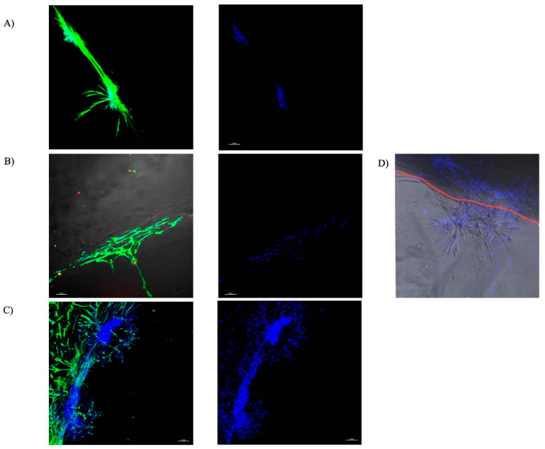
Figure 2.
Representative images of the ring/core interface with rat MSCs expressing α-SMA migrating into the core Gtn–HPA. DAPI, blue; Anti α-SMA, green (Scale bar 100 μ). (A) Control Gtn–HPA gel. (B) Gtn–HPA incorporating PDGF-BB. (C) Gtn–HPA incorporating PRP lysate. (D) representative image illustrating the superimposed image from the confocal microscopy of one of the samples showing the chain migration and the branching of the chains as the cells migrate into the gel. The red line illustrates the demarcation of the interface between the collagen ring and the core-Gtn–HPA hydrogel.

