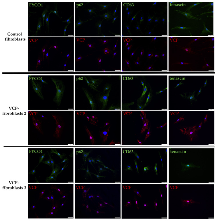
Figure 2.
Immunofluorescence studies on VCP–mutant and control fibroblasts with co–staining of VCP (red) and FYCO1, p62, CD63 and tenascin (green). VCP is increased in the cytoplasm and nuclei of patient derived fibroblasts. CD63 showed a strong cytoplasmic increase in patient fibroblasts compared to controls. FYCO1 and p62 were also more pronounced in patient fibroblasts. Tenascin is reduced in patient–derived fibroblasts. Scale bar = 50 µm. Two representative patients are shown: VCP fibroblasts 2 = patient 2 and VCP fibroblasts 3 = patient 3; patient numbering is consistent with patient numbering in Figure 1.

