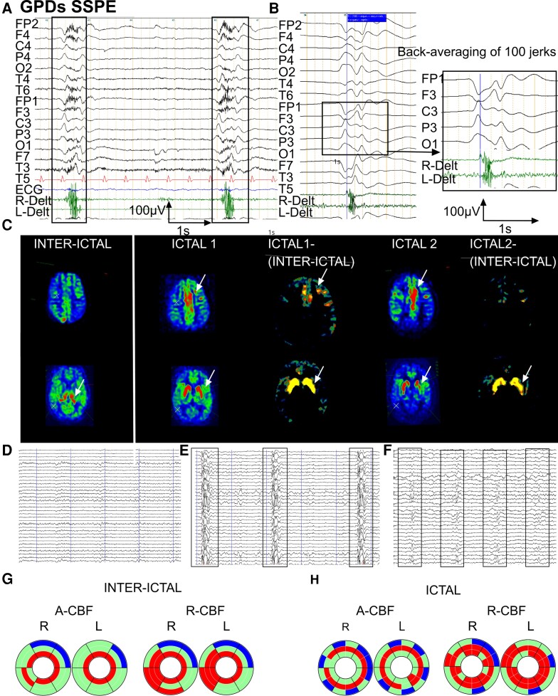
Figure 2.
Inter-ictal and ictal EEG-ASL-MRI in Patient 1, 15 years old, SSPE. (A) EEG with surface EMG polygraphic recording. Common average reference montage. Periodic diffuse high-amplitude slow-wave complexes of stereotyped morphology with maximum amplitude on left central-parietal leads occurring on a depressed background activity. Surface EMGs (deltoid muscles) of the concomitant jerks show a bilateral contraction beginning and predominating on the right deltoid. (B) Jerk-locked back-averaging (100 jerks), common average reference montage. Bilateral negative central-parietal sharp wave predominating on the left, reaching a maximal amplitude of 90µV with phase reversal over frontal areas. (C) ASL-MRI imaging in inter-ictal and ictal state [two examinations: ictal1 (1b) and ictal2 (1c) at 7 months interval] with ictal-(inter-ictal) subtraction. Arrows indicate structures with significant CBF increase. Compared with the inter-ictal state, ictal CBF was increased in frontal mesial and left frontal cortex. CBF in thalamus was increased in the inter-ictal state, whereas CBF here was normal in both adjacent cortex and in striatum. In ictal1 and ictal2 examinations, striatum showed increased CBF, while here CBF was unaffected in thalamus. (D) Simultaneous EEG recording during the ASL-MRI examination (1a). Slow and depressed background activity, neither GPDs nor jerks present. (E) Simultaneous EEG recording during the ASL-MRI examination (1b). GPDs concomitant with jerks on depressed background activity with complex-to-complex intervals of 5–7 s. (F) Simultaneous EEG recording during the ASL-MRI examination (1c). GPDs concomitant with jerks on depressed background activity showing shorter complex-to-complex intervals with further clinical evolution, of 2–3 s. (G) Donut charts (for details see Fig. 1B). Significant A-CBF and R-CBF changes compared with controls during inter-ictal state. A-CBF increased in all but one thalamic subdivisions and the right occipital striatum. R-CBF increased in all thalamic compartments as well as in bilateral temporal striatum, and in bilateral occipital C-S-T circuits. (H) Donut charts (for details see Fig. 1B and Supplementary Fig. 2). Significant A-CBF and R-CBF changes compared with controls during inter-ictal and both ictal examinations. A-CBF increased in all but two striatal subdivisions and in left caudal motor thalamus. R-CBF increased in all striatal and in most thalamic subdivisions, as well as in temporal cortical compartments.

