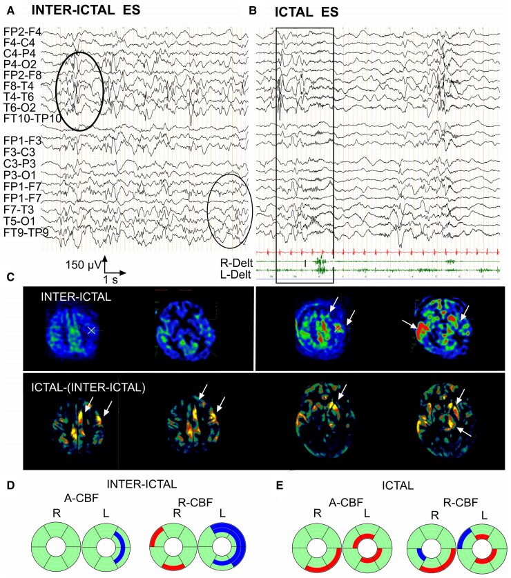
Figure 3.
Inter-ictal and ictal EEG-ASL-MRI in Patient 26, 6 months old, epileptic spasms/West syndrome with bilateral extended cortical dysplasia. (A) Inter-ictal EEG, awake. Independent slow-wave and spike-wave foci in left and right central-parietal-temporal regions. (B) Ictal EEG with surface EMG polygraphic recording, bipolar montage. Asymmetric epileptic spasm predominating on the right deltoid muscle concomitant on EEG with a diffuse slow-wave complex associated with low-voltage, diffuse fast rhythm bursts predominating on left parietal-temporal region. (C) ASL-MRI imaging in inter-ictal and ictal state with ictal-(inter-ictal) subtraction. Arrows indicate structures with significant CBF increase. Ictal CBF is increased in central-mesial and left frontal cortex compared with their inter-ictal CBF. Ictal CBF was also increased in the left striatum, left thalamus and right temporal cortex compared with their inter-ictal CBF. (D) Donut charts (for details, see Fig. 1B and Supplementary Fig. 3). Significant A-CBF and R-CBF changes compared with controls during inter-ictal state. A-CBF decreased in two right S (rostral motor, caudal motor). R-CBF increased in two right C (parietal, temporal) and decreased in three corresponding left C and S (prefrontal, rostral motor, caudal motor) and in left parietal S. (E) Donut charts (for details, see Fig. 1B and Supplementary Fig. 3). Significant A-CBF and R-CBF changes compared with controls during ictal state. A-CBF increased in four left T (prefrontal, caudal motor, parietal and temporal) and in two right C (caudal motor, parietal). R-CBF increased in three left T (prefrontal, caudal motor, parietal) and two right-sided C (parietal, caudal motor).

