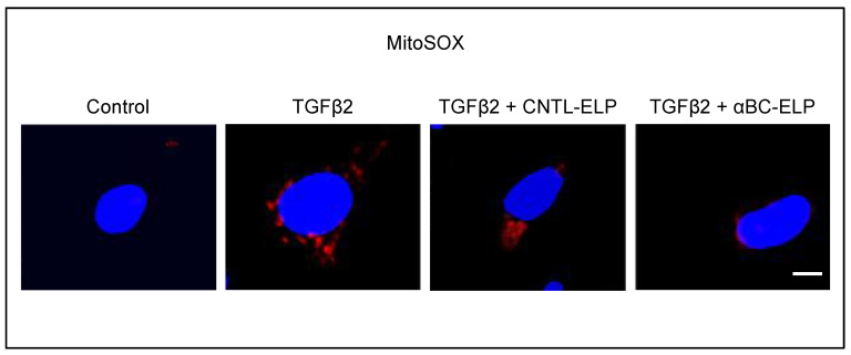
Figure 3.
Effect of TGFβ2 on ROS generation and inhibition of ROS by αBC-ELP. hRPE cells were cultured in 4 well chamber slides. Twenty four hour after treatment with TGFβ2 (10 ng/mL) or co-treatment with TGFβ2 (10 ng/mL) and αBC-ELP or co-treatment with TGFβ2 (10 ng/mL) and CNTL-ELP, cells were incubated with 5 µM MitoSox Red (mitochondrial superoxide marker) for 10 min at 37 °C. After washing, cells were imaged with a confocal microscope (ZEISS LSM 710).TGFβ2 at 10 ng/mL (24 h) noticeably increased mitochondrial ROS production in RPE cells and co-treatment with αBC-ELP at 10 µM inhibited TGFβ2 -induced ROS formation. SI is used as negative control (CNTL-ELP). Red: Mitochondrial superoxide, Blue, DAPI, nuclear stain. Scale bar: 10 µm.

