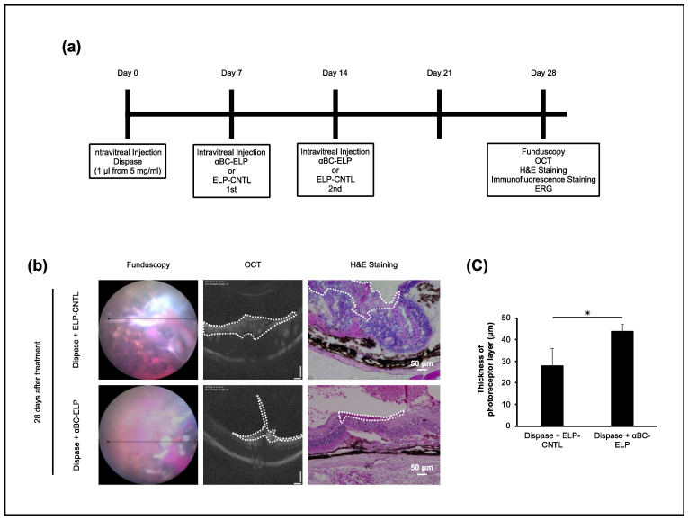
Figure 8.
Effect of αBC-ELP in dispase-induced PVR in mouse. Mice were injected with a single intravitreal dose of dispase (1 μL, 5 mg/mL) on day 0. On days 7 and 14, αBC-ELP (CrySI, 1 μL from 218 μM) or Control ELP (SI) was administered intravitreally. On day 28, fundus, OCT images, and ERG data were gathered. At the end of the procedure, mice were euthanized, and retinal sections were processed for H&E and immunostaining. (a) Experimental scheme. The ELP preparations for α-BC-ELP and ELP-CNTL are also referred as CrySI and SI, respectively. (b) Fundus, OCT images, and H&E staining showed that mice treated with αBC-ELP had less proliferative membrane area and decreased retinal layer structure disruption compared to the dispase + ELP-CNTL group (b) The area enclosed by the white dotted line in OCT and H&E staining represents the PVR membrane. (c) Quantification of the photoreceptor thickness. αBC-ELP: αB crystallin-Elastin-like peptide. Scale bar: 50 μm. * p < 0.05, n = 5.

