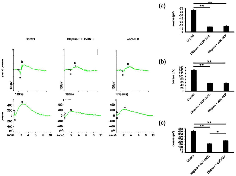
Figure 9.
Effect of αBC-ELP on photoreceptor function by ERG. The experimental protocol is shown in Figure 8a. Electroretinogram (ERG) analysis of mice treated with and without αBC-ELP (a–c). The ‘a’—wave represents the hyperpolarization of photoreceptors (a), the ‘b’ wave represents the second-order neuron response (b), and the ‘c’ wave represents light-induced activity in the photoreceptors (c). Three groups were compared: (1) wild-type with PBS as control, (2) dispase (1 μL, 5 mg/mL) + ELP-CNTL treated, (3) dispase (1 μL, 5 mg/mL) + αBC-ELP (1 μL from 218 μM). While there is no change in (a,b) waves, (c) wave amplitude significantly improved with αBC-ELP. Values are the means ± SE. αBC-ELP: αB crystallin-elastin-like polypeptide. * p < 0.05, ** p < 0.005. n = 3.

