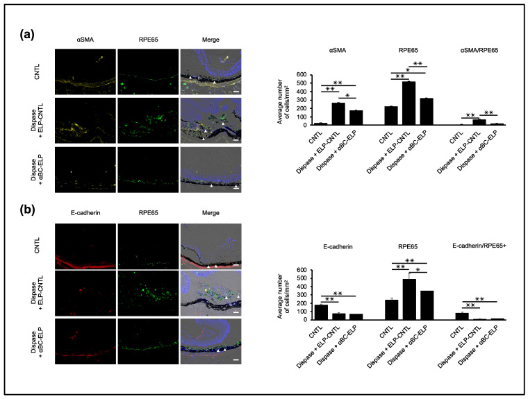
Figure 10.
αBC-ELP inhibits EMT in dispase-induced PVR in mice. The experimental protocol is shown in Figure 8a. Double immunofluorescence staining for αSMA or E-cadherin with RPE65 in retinal sections (a,b). Nuclei are stained blue. White arrowheads indicate co-staining of αSMA/RPE65 and E-cadherin/RPE65, respectively. Quantification of number of positive cells for αSMA, RPE65, E-Cadherin and co-localization is shown in the bar graphs. Values are means ± SEM. N.S.: not significant, αBC-ELP: αB crystallin-Elastin-like peptide. Scale bar: 50 μm. * p < 0.05, ** p < 0.01. n = 10.

