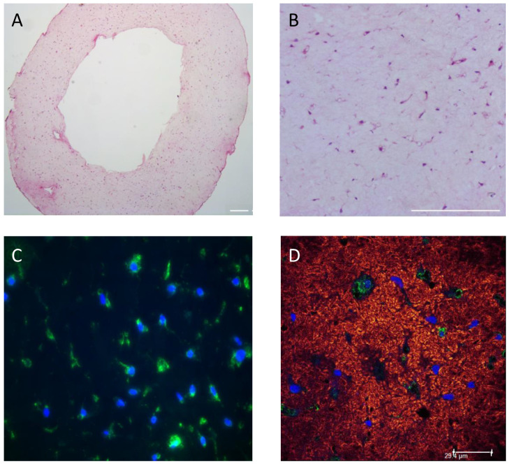
Figure 4.
Histological and immunohistological analysis of EHM-ring sections. HE staining of an EHM ring section showing even distribution of the cells throughout the EHM ring structure. EHM ring was formed using 1.5 mg/mL collagen concentration and 1000 CF cells/μL gel and was fixed and stained 24 h after casting. Darker dots implicate CF. Magnification 5× (A); or 20× (B). Bars represent 200 µm. Vimentin (green) and DAPI (blue) staining (C); and Z-stack image of CNA35 (red) and Vimentin (green) staining nuclei are blue (DAPI) (D) (bar represents 29.4 μm).

