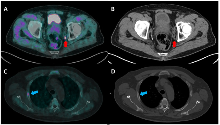
Figure 2.
(A,B) A 71-year-old man with locally advanced PCa (Gleason score 9, 4 + 5 and a serum PSA value of 36 ng/mL) underwent [18F]F-choline PET/CT ((A) transaxial fused image; (B) CT image) for the initial staging of the disease. The PET/CT image shows a pathological uptake corresponding to a small left internal iliac lymph node (red arrow). Lymph node metastasis was not detected in CT staging, as its size (0.7 × 0.6 mm) was below the significance criteria. (C,D) A 68 year-old-man with PCa (G.S 6, 3 + 3; PSA value of 71.29 ng/mL) was imaged with [18F]F-choline PET/CT staging ((C) transaxial fused image; (D) CT image), with evidence of a focal uptake corresponding to a bone lesion at the third right rib (blue arrow), which could not be detected by CT due to the normal bone structure.

