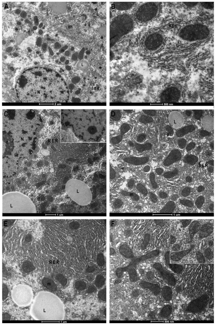
Figure 4.
Transmission Electron Microscopy micrographs of control and BTBR mice liver. Control (CTR) (A,B) and BTBR mice liver (C–F). CTR liver shows regular nucleus (N), few lipids drop (L), well-preserved mitochondria (m) associated with rough (RER) or smooth reticulum (SER) (A). Greater magnification of mitochondria, surrounded by RER (B). BTBR liver shows irregular nuclear envelope, hypertrophic nucleoli and RER with congested cisterns, increased number and size of lipid drops (C). Mitochondria, mostly small in size (C–E) and mitochondrial fission phenomena ((F) and insert) are recognizable. Asterisk: bile canaliculus; (g) glycogen; (arrow) nuclear pore. Magnification: (A) 2550×, (B) 9900× (C) 4000×, (D) 7800×, (E) 7000×, (F) 9900×.

