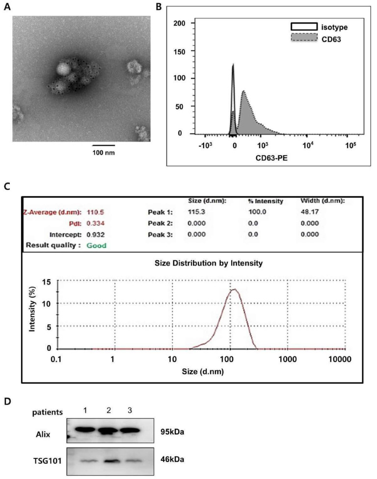
Figure 1.

Urinary exosome characterization. (A) CD63 immunogold stain image of exosomes. Representative transmission electron micrographs of exosomes labeled with gold particles (black dots) are shown. (B) Flow cytometry analysis of the exosomal CD63 marker. (C) Size distribution of urinary exosomes analyzed by Zeta sizer. (D) Western blot analysis of the expression of exosomal markers Alix and TSG101 in urinary exosome proteins. ABMR, antibody−mediated rejection; TCMR, T cell−mediated rejection; NOMOA, no major abnormality; BKVN, BK virus nephropathy. FC, fold change.
