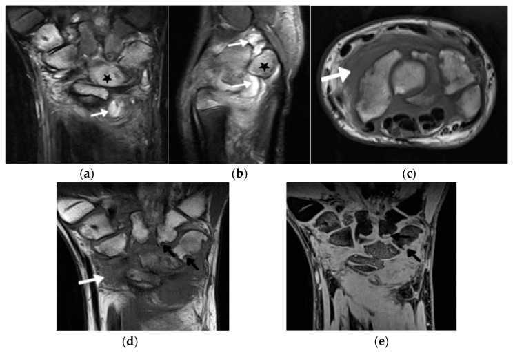
Figure 3.
A 14-year-old boy with JIA and wrist arthritis. Coronal T2 Turbo inversion recovery magnitude (TIRM) (a), sagittal proton density fat saturated (b) and axial T1-weighted time spin echo (TSE) (c) MR images show synovitis (white arrows) and bone marrow edema (stars); coronal T1 TSE (d) and T1 volume-interpolated breath-hold examination (VIBE) (e) show numerous cyst-like changes and erosions (black arrows).

