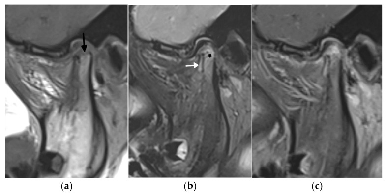
Figure 5.
A 1.5 Tesla temporomandibular joint MRI with T1- (a), fat-saturated T2- (b), and postcontrast fat-saturated T1-weighted images (c) in a 7-year-old girl with juvenile idiopathic arthritis showing active and chronic inflammatory lesions: synovitis (white arrow), bone marrow edema in the mandibular condyle (star) in the upper part of the ramus, and erosions (black arrow).

