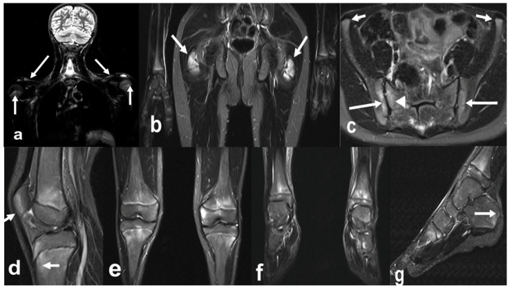
Figure 7.
Whole-body MRI (WBMRI) in a 12-year-old boy with enthesitis-related arthritis (ERA) subtype of juvenile idiopathic arthritis (JIA). Superposition of findings of ERA and chronic recurrent multifocal osteomyelitis (CRMO) is seen on this WBMRI examination with STIR images of multiple body parts in multiple directions (a–g). Findings of CRMO are presented by ill-defined flamed-shaped areas of increased inversion recovery (IR) signal seen in the bilateral proximal humeral metaphyses and distal clavicles (a, arrows), distal femoral and proximal tibial metaphyses and at a lesser extent, epiphyses (d,e), distal tibial metaphyses and epiphyses, and tarsal and proximal metatarsals (g). Findings of JIA-related enthesopathies are represented by IR signal noted in the bilateral greater trochanters (b, arrows) at the site of insertion of gluteus medius and minimus, along the sacroiliac joints (c), iliac, long arrows and sacral aspects, arrowhead; (c), normal anterior–superior iliac spine apophyses short arrows), proximal and distal attachments of patellar tendon (d, arrows), and at the insertion of the Achilles tendon in the posterior calcaneus (g, arrow).

