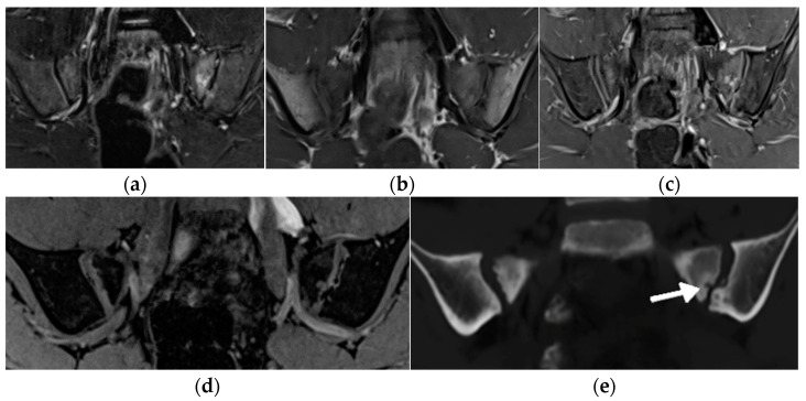
Figure 9.
MRI of the sacroiliac joints of a 11-year-old boy with an active erosion in the left sacrum seen in consecutive sequences: T1-weighted time spin echo (TSE) (a), T2 short tau inversion recovery (STIR) (b), postgadolinium fat-saturated T1-weighted MR image (c), T1 volume-interpolated breath-hold examination (VIBE) (d), and BoneMRI (e, arrow).

