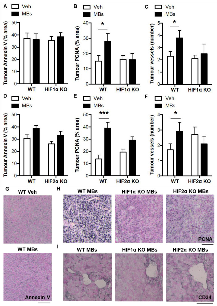
Figure 5.
Characterization of pulmonary tumors from vehicle- and microbead-treated WT and myeloid HIFα KO mice. (A) Pulmonary tumor apoptosis, (B) proliferation, and (C) vascularisation in pulmonary tumors following administration of intravenous vehicle (Veh) or microbeads (MBs) at 1 day post-LLCs in WT mice and myeloid HIF1α KO littermates. (D) Pulmonary tumor apoptosis, (E) proliferation, and (F) vascularisation in pulmonary tumors following administration of intravenous vehicle (Veh) or microbeads (MBs) at 1 day post-LLCs in WT mice and myeloid HIF1α KO littermates. Panels (A,B,D,E) are quantifications of the percentage area of the pulmonary tumor that was occupied by positive staining for annexin V (apoptosis) or PCNA (proliferation). Quantifications were performed by image analysis. In panels C and F, the number of tumor vessels was counter using a Chalkley reticule and normalized to tumor surface area. (G) Representative images of pulmonary tumor sections with immunostaining (black) for Annexin V, (H) PCNA, and (I) CD34. Scale bar = 50 μm. Mice were culled and lung tissue excised for analysis at day 14 post-LLCs. N = 10/group. * p < 0.05 and *** p < 0.001.

