Figure 2.
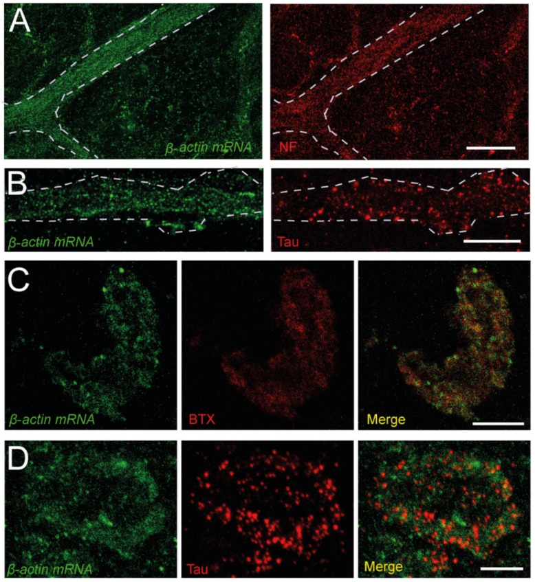
β-actin mRNA is present in peripheral axons and NMJs. (A). Example of β-actin mRNA (green) in a bundle of axons visualized by FISH (left panel). The same axons are marked with an anti-NF antibody (red, right panel). (B). Detail of the dotted signal of the probe in a band of axons (left panel), identified by its immunoreactivity to Tau (red, right panel). (C). Images of an NMJ displaying β-actin mRNA (green, left panel) and BTX (red, middle panel), and a merged image (right panel) (D). Images of an NMJ displaying β-actin mRNA (green, left panel) and Tau (red, middle panel) signals, and a merged image (right panel). Calibration bars: (A) 40 µm, (B–D) 10 µm.

