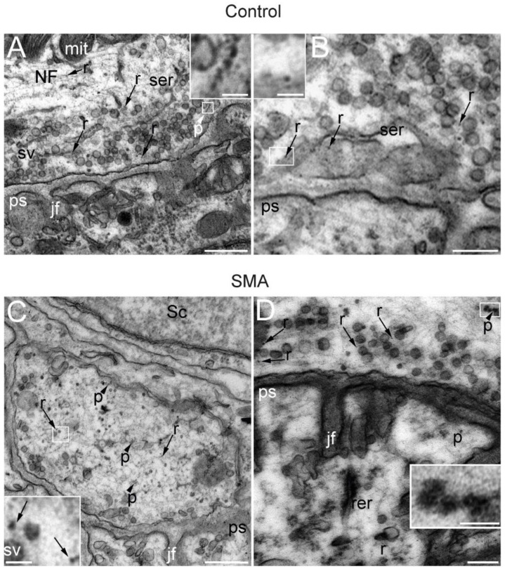
Figure 3.
SMA and control presynaptic motor terminals contain ribosomes and polysomes. Electron microscopy representative images of NMJs from the TA muscle of control (A,B) and SMA (C,D) mice from the SMNΔ7 line at P14. In the cytoplasm of the presynaptic terminal, synaptic vesicles (sv), neurofilaments (NFs), mitochondria (mit), and smooth endoplasmic reticulum (ser) are indicated. Scattered throughout the cytoplasm of the presynaptic terminal are numerous independent ribosomes (r) (arrows and boxes in B,C) and, less abundantly, polysomes (p) (arrowheads and boxes in A,D). Other identified elements incude jf: junction folds; ps: postsynaptic element; rer: rough endoplasmic reticulum; Sc: Schwann cell. Calibration bars: 500 nm (A,C), 200 nm (B,D), and 50 nm (inserts from A,D).

