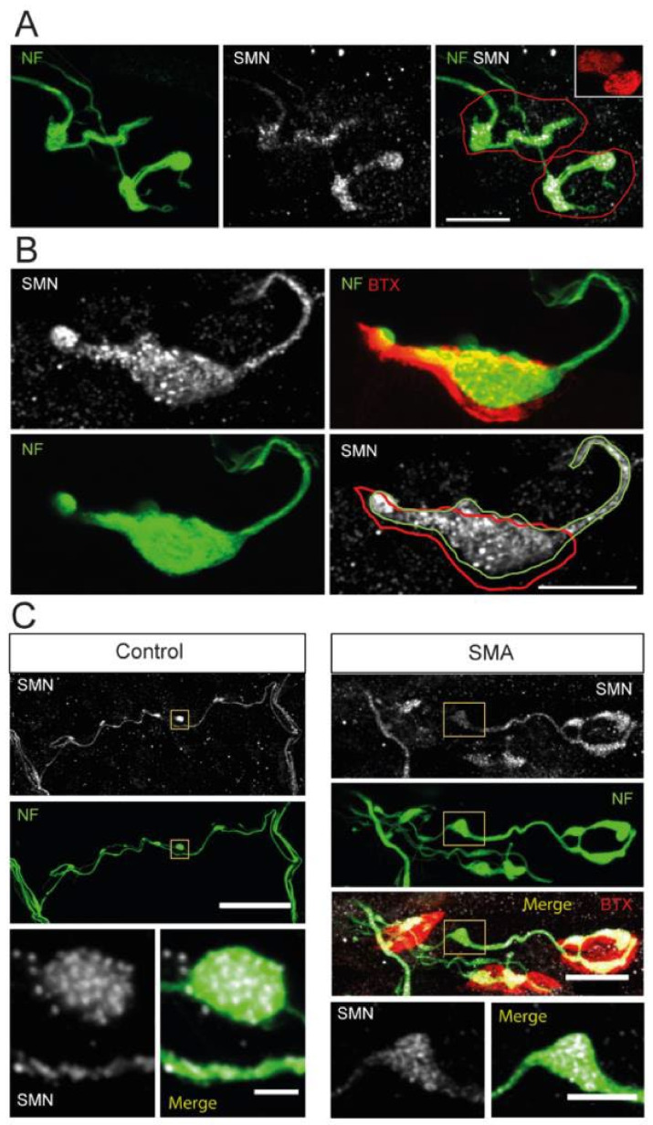
Figure 8.
SMN aggregates in NF accumulations at P9. (A). Representative example of motor synaptic terminals (red outlines) displaying few NFs branches (green) and aggregates of SMN granules (white) in an SMA mouse. Scale bar: 10 µm. (B). SMA presynaptic terminal showing a massive accumulation of NFs (outlined in green), occupying much of the terminal area (red outline) and containing abundant SMN granules (white). Scale bar: 10 µm. (C). Axonal accumulations of NFs in the SMNΔ7 model coinciding with SMN granule aggregates, both in transgenic control and SMA mice. The insets show details of the overlap of SMN granules and NFs. Accumulations of SMN granules and NFs were only occasionally observed in control mice. The scale bars are 40 and 2 µm (left panels) and 15 and 5 µm (right panels).

