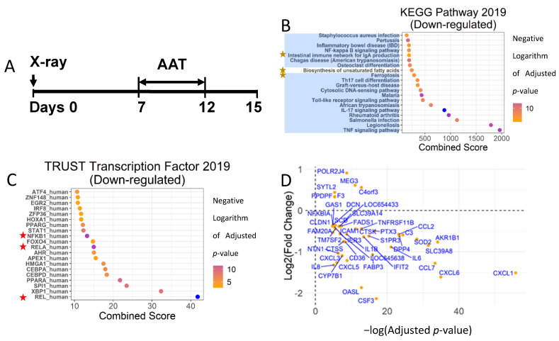
Figure 1.
Human AAT suppressed NF-κB-mediated inflammatory pathways in senescent HCA2 cells. (A) Study strategy: the HCA2 cells were irradiated and cultured without any interference for 7 days and then treated with hAAT (2 mg/mL) or 1× PBS containing fresh medium for 5 days. At day 12, FBS-free medium was used to replace the hAAT- or PBS-containing medium and culture the cells for 3 days. Total RNA was extracted at day 12 or 15 and processed for RNA-seq or PCR. (B) Pathway enrichment analysis using KEGG Pathway 2019 Gene Set. Blue shade: pathways related to inflammation; yellow star: pathways not regulated by NF-κB. (C) Transcription factor enrichment analysis using TRUST Transcription Factor 2019 Gene Set. Red star: NF-κB subunits. (D) The DEGs were identified by an adjusted p-value less than 0.01. The log2 (fold change) of DEGs were plotted against-log (adjusted p-value). The combined score was the combination of the Fisher’s exact test p-value and deviation from the expected rank of each term. The p-value, adjusted p-value, and combined score were calculated following [30].

