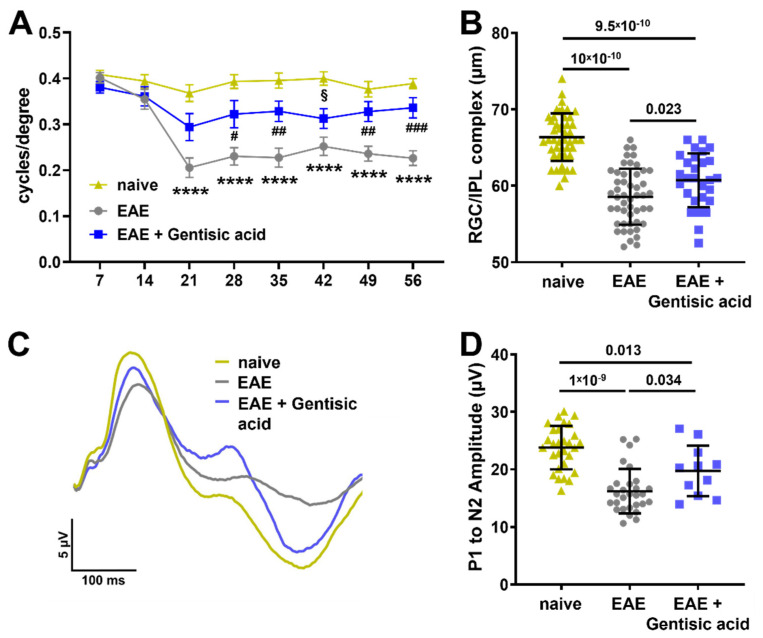
Figure 4.
Gentisic acid (GA) treatment in EAE animals improves visual function and structure: (A) assessment of visual acuity over time demonstrates a significant decrease in both untreated EAE mice and GA-treated EAE animals as of 21 days after EAE induction. More importantly, EAE mice in the GA treatment cohort show a solid preservation in visual acuity thereafter with a trend towards recovery. Mean data are given with SEM, **** p < 0.0001 in EAE vs. naïve, # p < 0.05, ## p < 0.01, ### p < 0.001 in EAE vs. EAE + GA, and § p < 0.05 in EAE + GA vs. naïve. (B) Analysis of the GCL/IPL complex using OCT indicates a significant decrease in thickness in untreated EAE mice whereas GA treatment significantly ameliorates GCL/IPL complex thinning. (C) Cumulative pattern ERG tracing results from each group demonstrate a clear reduction in the amplitudes in untreated EAE mice and a preserved pattern ERG waveform in EAE animals having received GA. (D) The P1-N2 amplitude in EAE mice is significantly reduced when compared to GA-treated EAE animals as well as naïve mice. Data shown in (B) and (D) represent individual eyes with mean ± SD.

