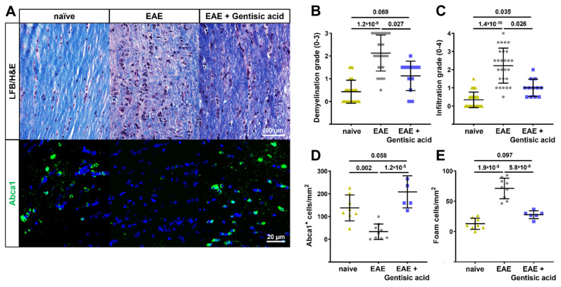
Figure 5.
Optic nerve histopathology in EAE mice. (A) Representative images of longitudinal optic nerve sections stained with LFB/H&E and Abca1 immunohistochemistry. (B) Gentisic acid (GA) treatment of EAE mice preserved the myelin sheath when compared to untreated EAE animals, as indicated by ordinal gradings of demyelination. (C) Similarly, EAE mice treated with GA demonstrated reduced cell infiltration whereas untreated EAE mice showed massive grades of infiltration, which is an indication of active optic neuritis. (D) Furthermore, systemic GA administration rescued the number of Abca1+ cells present and significantly reduced the number of foam cells (E) in EAE optic nerves. All data are given as mean ± SD.

