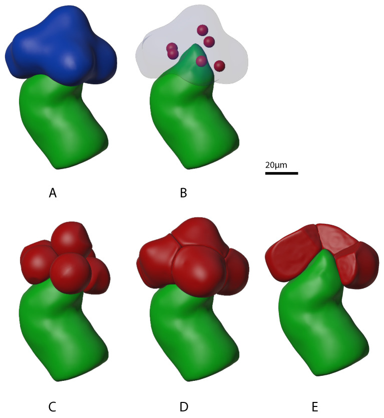
Figure 4.
Progression of mini-gland individual acinus outer boundary “growth” stages. The mini-gland duct cell outer boundary from our prior work [27] is shown in green. (Panel A) shows the mini-gland acini outer boundary in blue. (Panel B) shows the arrangement of six individual acinus “seeds” in red. (Panel C) shows partially grown individual acini and (Panel D) shows the fully grown acini. (Panel E) shows the mini-gland with several of the acinus outer boundaries (red) removed. Note that the acini are packed tightly against each other as well as to the tip of the mini-gland duct outer boundary.

