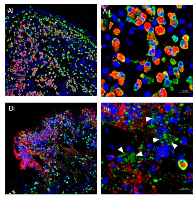
Figure 2.
(A,B) Immunofluorescence staining of nasal polyps from patients with chronic rhinosinusitis with nasal polyps. Fluorescence images for MBP (red), galectin-10 (green), and DNA (blue) were obtained using a Carl Zeiss LSM780 confocal microscope, as previously described [36]. Intact eosinophils are massively infiltrated in polyps (Ai,Aii). The presence of ETotic eosinophils with extracellular traps (degenerated DNA), cell-free granules (punctate MBP), extracellular vesicles (punctate galectin-10), and CLCs (galectin-10-stained needle-like crystals; arrowheads) can be seen (Bi,Bii). Scale bars: 50 μm for (i) and 10 μm for (ii).

