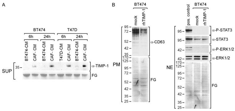
Figure 6.
CAF-CM leads to an increase in exposure of BT474 and T47D cells to TIMP-1. (A) Western blot analysis of the supernatants (SUP) from BT474 and T47D cells after treatment with BT474-CM, T47D-CM or CAF-CM for the amounts of time as indicated. (B) Western blot analysis of plasma membrane extracts (PM) or nuclear extract (NE) from BT474 cells for the proteins and phospho-proteins as indicated. To show equal protein loading, the membranes were stained by Fast Green (FG).

