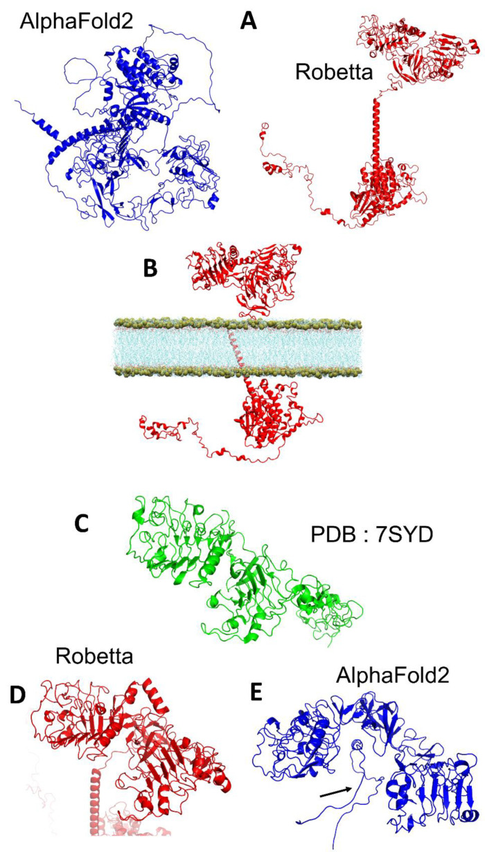
Figure 1.
Comparison of the structure of the epidermal growth factor receptor retrieved from Alpha Fold (depicted as cartoon colored in blue) or modelized via Ab-initio calculation on Robetta (red) (A). Molecular model of the insertion of the receptor obtained by Robetta in a lipid membrane environment (B). Comparison of the structure of the epidermal growth factor receptor resolved by Cryo-EM (PDB: 7SYD, resolved from the residue 25 to 638) ((C), green) with the structure obtained by Robetta ((D), red) and AlphaFold2 ((E), blue). In (E), the arrow points to an unstructured region that was predicted and inserted by the AlphaFold2 algorithm between the two extra-cellular domains.

