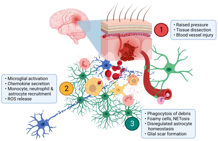
Figure 1.
Pathophysiology of ICH. (1) Haemorrhage from a parenchymal arteriole drives pressure gradients that dissect neural tissue (blue) and may cause distant injury as well as blood vessel injury. (2) Myelomononuclear cells (yellow) rapidly respond by release of proinflammatory cytokines and other chemokines. These serve to recruit MdCs, astrocytes (green) and neutrophils (amber) which secrete further inflammatory mediators and reactive oxygen species (ROS). (3) As inflammation progresses, debris is progressively phagocytosed and foamy myelomononuclear cells appear. Neutrophil NETosis may serve to limit haemorrhage and/or microvascular blood flow. As astrocytes respond to the ICH and contribute to glial scar formation, their homeostatic functions including neurovascular and neurometabolic coupling are impaired. Created with BioRender.com.

