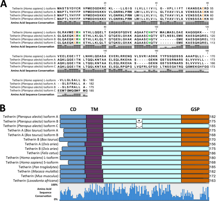
FIG 3.
Multiple sequence alignment (MSA) and protein domains of mammalian tetherin amino acid sequences. (A) MSA reveals amino acid residues differing between human and P. alecto sequences, indicated by the amino acid sequence conservation bar graph below the sequences. Conserved cysteine and asparagine residues are colored orange and green, respectively. (B) Protein domains of mammalian tetherin proteins are depicted as overlays of an MSA. Sequence conservation is indicated by the bar graph below the alignment. Protein domains are color coded: CD, cytoplasmic domain (blue); TM, transmembrane domain (purple); ED, extracellular domain (light blue); GSP, glycophosphatidylinositol signal peptide (orange). Asterisk (*) indicates a 7-amino acid (aa) exclusion in P. alecto tetherin isoforms B and C relative to isoform A.

