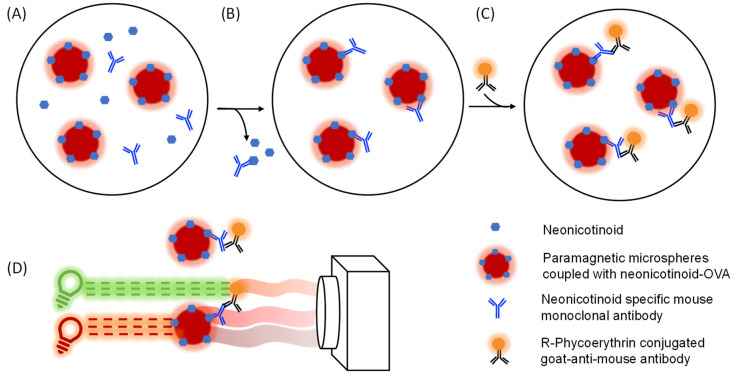
Figure 2.
Assay and detection principle of the neonicotinoid microsphere immunoassays (nMIAs). OVA–neonicotinoid conjugates that are coupled microspheres are incubated with specific neonicotinoid antibodies and a sample containing free neonicotinoids. The OVA–neonicotinoid conjugates and free neonicotinoids compete in antibody binding (A). After incubation, the unbound antibodies and neonicotinoids are washed away (B). When it is incubated with an uncontaminated sample, the neonicotinoid antibody binds to the OVA–neonicotinoid conjugates on the microspheres, while fewer antibodies are bound to the microsphere when they are incubated with a neonicotinoid-contaminated sample. Next, an ample number of secondary anti-species antibodies that are coupled to R-phycoerythrin are incubated with the OVA-neonicotinoid-antibody complex and the excess is washed away (C). The microspheres contain two unique fluorochromes which emit red and far-red light upon excitation by a red LED. These emissions are captured using the CCD camera in the planar array reader for microsphere identification. Next, the intensity of the orange-emitted light from the reporter is measured after excitation by the green LED (D).

