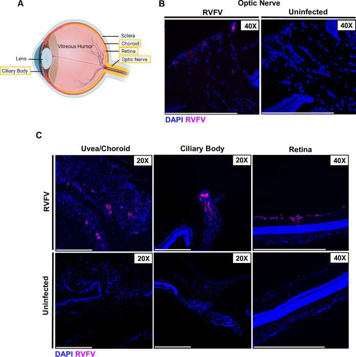
FIG 3.
Tissue tropism of RVFV in the posterior portion of the eye at 3 dpi. (A) Schematic of ocular anatomy with structures of interest boxed in yellow. Created in BioRender.com. (B) Eyes were enucleated from Sprague Dawley rats 3 days following subcutaneous RVFV infection. RVFV antigen staining was found on the optic nerve head using an anti-RVFV N antibody (magenta). (C) RVFV was also found in the uvea, ciliary body, and ganglion layer of the retina at 3 dpi. Scale bar is 250 μm.

