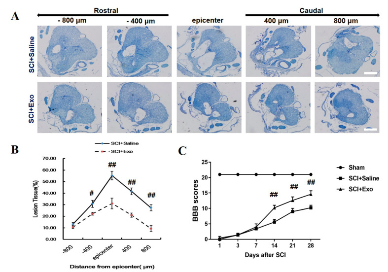
Figure 2.
M2 macrophage exosomes attenuate TD and improve FR post-SCI. Representative cresyl violet staining of the SC lesion cavity (A). Quantification of the lesion cavity; the ratio presents as ‘‘lesion area/total area’’, 800 µm caudal and rostral to the epicenter, after 28 days of SCI (B). FR of rats was accessed by BBB scores and ranged from day 1 to day 28 (C). Mean ± SD. # p < 0.05 SCI + Exo versus PBS group, ## p < 0.01 SCI + Exo versus PBS group. n = 5 per group. Scale bar = 1 mm.

