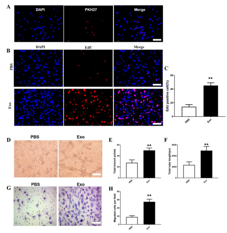
Figure 6.
M2 macrophage exosomes attenuate angiogenesis, migration, and proliferation of bEnd.3 cells. PKH27-labelled exosomes uptake by bEnd.3 cells. Scale bar = 50 µm (A). The proliferation of HUVECs in each group (B). Quantification of EdU+ cells in each group (C). Tube formations were measured after growing bEnd.3 cells pre-treated with exosomes or PBS. Scale bar = 500 µm (D–F). Migration areas were measured in each group in bEnd.3 cells (G). Quantitative analysis of the numbers of the migrated bEnd.3 cells (H). Mean ± SD. ** p < 0.01 SCI + Exo versus PBS group. N = 5 per group.

