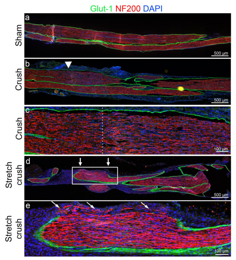
Figure 3.
Histological comparison of acute six-day sciatic nerve injuries. (a) One-week uninjured sham nerve. (b) Crush injured nerve—note intact perineurium (Glut1, a marker of perineurium); yet, increased cellularity and potential inflammation related to Wallerian degeneration at and just beyond the crush site (white arrowhead). (c) Magnified crush-injury site of (b), dotted line marks the crush epicenter. Note again intact perineurium and the intense localization of nuclei (DAPI) distal (right of dotted line) to the crush site. In addition, thinned out disarrayed axons, compared to proximal to the crush zone, are evident. (d) Stretch–crush injured nerve. Note perineurium disruption at the injury site (between white arrows), significant nerve swelling and increased cellularity supporting an NIC development. (e) Magnified stretch–crush injury of (d), showing perineurial disruption as seen by Glut1 disruption, disarrayed axonal architecture with increased cellularity and several axons evident beyond the perineurial border—forming an NIC. NIC, neuroma in continuity.

