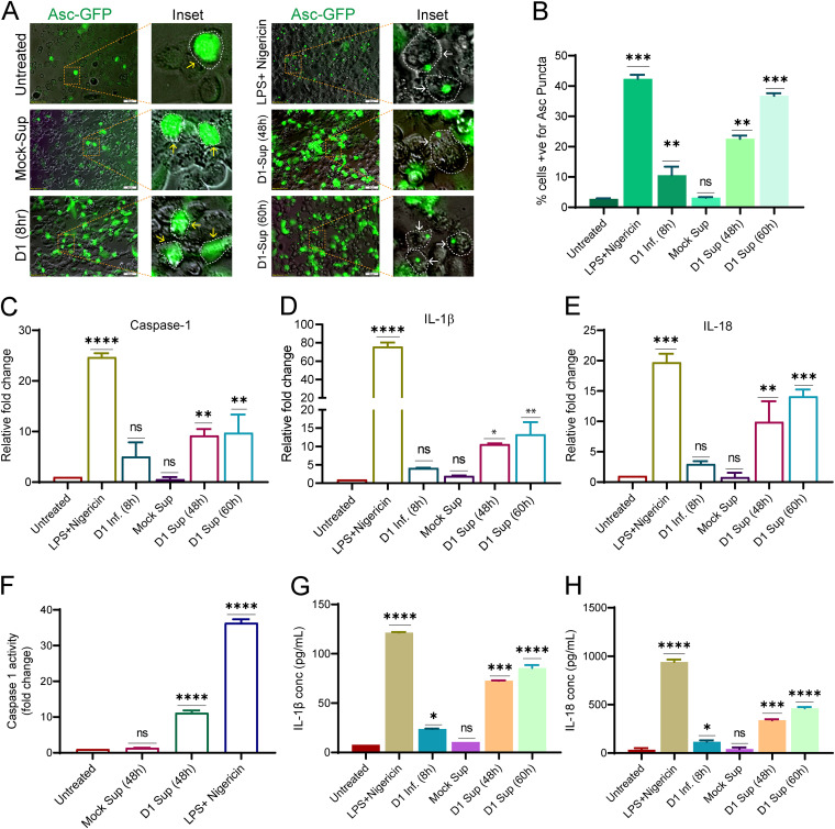
FIG 9.
Dengue infection releases mt-DAMPs, which leads to inflammasome activation in monocytes and macrophages. (A) The inflammasome activation reporter monocyte cell line THP-ASC-GFP-derived macrophages, were challenged for 8 h with preclarified culture supernatants obtained from DENV-infected (serotype 1)-Huh7 cells at 48 h and 60 h postinfection. Challenge with culture supernatant from mock-infected Huh7 cells, LPS+Nigericin, and direct infection with dengue virus 1 was used as the negative, positive, and infection control. The upregulation of ASC-GFP expression (inflammasome priming) and ASC-GFP specks formation is represented by yellow and white arrows in the respective zoomed insets. The cell periphery is indicated by dotted lines. (B) Bar graph depicting the quantification of the number of ASC-GFP puncta/specks per cell 8 h postchallenge with the respective treatments. (C to E) Graphs depicting the transcript levels of caspase 1 (C), IL-1β (D), and IL-18 (E), in THP1 monocytes challenged for 8 h with the respective treatments described in panel A. The data are presented as the fold change with respect to untreated THP1 monocytes. (F) Graph depicting the relative caspase 1 activity in THP1 monocytes 8 h postchallenge with the indicated treatments. (G and H) Graph depicting the relative quantity of IL-1β and IL-18 released into the culture supernatants from the activated THP1 monocytes 8 h postchallenge with the treatments indicated in panel A. Data are the mean ± SEM from three independent experiments. Statistical significance was determined using one-way ANOVA. ns, nonsignificant; *, P < 0.05; **, P < 0.01; ***, P < 0.001, ****, P < 0.0001.

