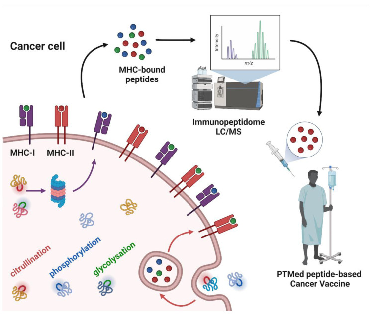
Figure 1.
Post-translationally modified peptide-based cancer vaccine workflow. The figure depicts cancer cell antigen processing of intracellular and extracellular proteins, subsequently as peptides bound to MHC-I or MHC-II. Some of the proteins have PTMs in their structure, which are sketched in colors (citrullination: red; phosphorylation: blue; glycosylation: green) as well as in MHC-bound peptides. The MHC-bound peptides are identified by means of liquid chromatography-mass spectrometry (LC/MS), to derive the cancer cell immunopeptidome. From the immunopeptidome data, peptides with PTMs can be selected as antigens for cancer vaccines.

