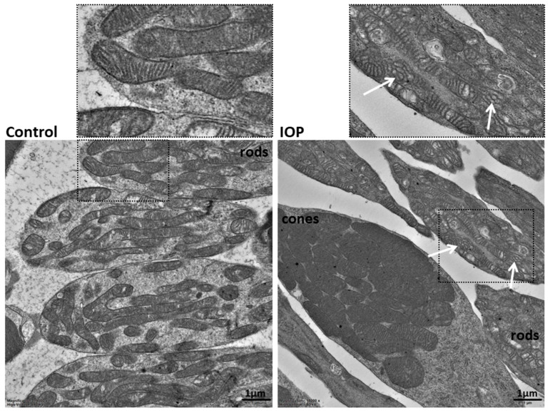
Figure 2.
Ultrastructural features of the mitochondria in the rods of the retinal ischemia-induced (right image) and control (left image) eye. The cristae of the mitochondria are compact and narrow in the control eye. White arrows show the presence of dilated cristae with abnormal shapes in the mitochondria of the high-pressure-induced retinal ischemia model. Two ultrathin sections of the neuroretina were observed, 64 RI cells and 56 control cells were used to evaluate the mitochondrial phenotype in rods, and the phenotype appeared homogeneous throughout the sample. TEM, 15,000×.

