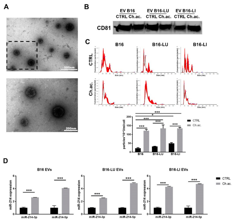
Figure 1.
Extracellular vesicles released by melanoma cells. (A) Transmission electron microscopy (TEM) of B16-derived EV at different magnifications (upper picture: scale bar = 500 nm; lower picture: scale bar = 200 nm, magnification of the squared area above). (B) Western blot analysis of CD81 of melanoma-derived EV. (C) Nanoparticle tracking analysis (NTA) at Nanosight NS300 of EV released by B16, B16-LU, and B16-LI cells under standard (CTRL) and chronic extracellular acidosis (Ch.ac.) (one-way ANOVA). (D) miR-214-3p and miR-214-5p expression levels in EV released by B16, B16-LU, and B16-LI under standard and chronic acidic conditions (two-way ANOVA). * p < 0.05 and *** p < 0.001.

