Figure 2.
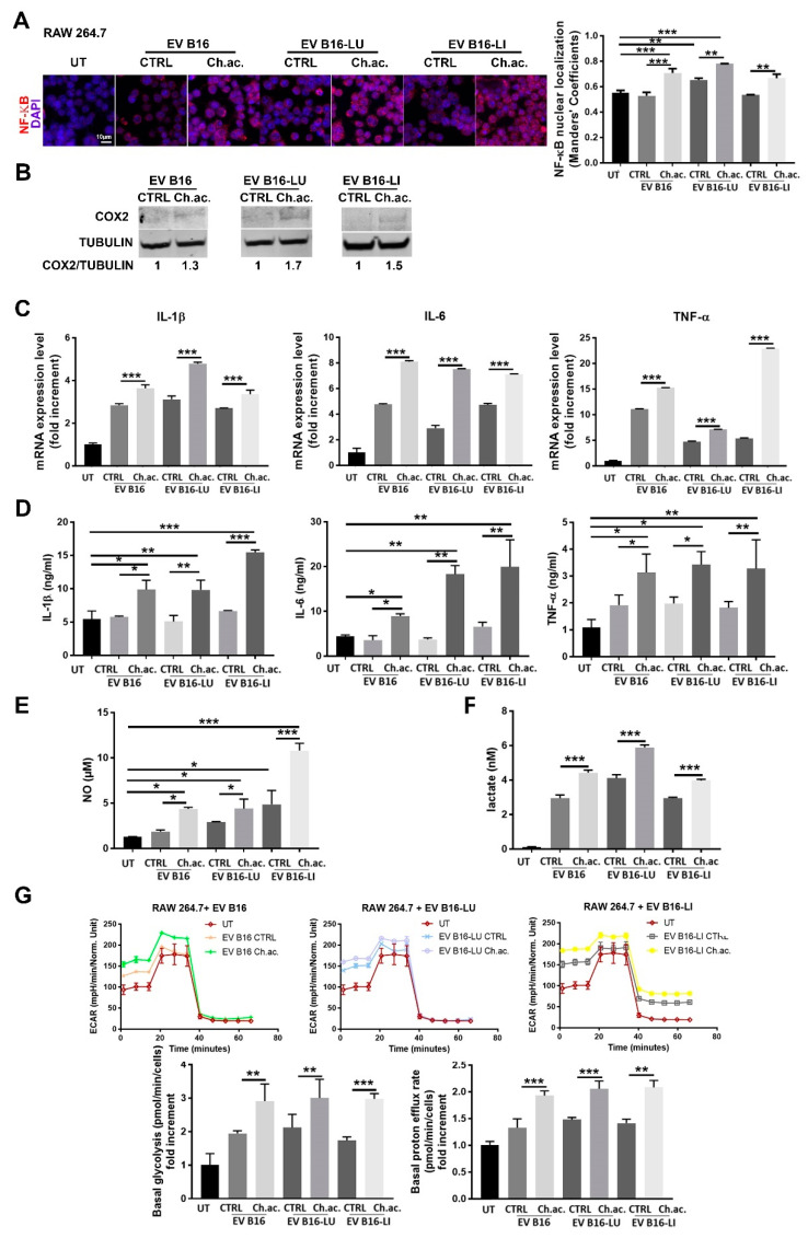
Inflammatory response of RAW 264.7 macrophages following the uptake of melanoma-derived acid-EV. (A) Immunofluorescence analysis of NF-ĸB nuclear localization in RAW 264.7 macrophages treated with melanoma-derived EV (Scale bar = 10 µm). (B) Western blot analysis of COX-2 in RAW 264.7 macrophages treated with melanoma-derived EV (p < 0.05). (C) Real-time qPCR of IL-1β, IL-6, and TNF-α in RAW 264.7 macrophages treated with melanoma-derived EV (all conditions were found significantly different from the untreated (UT). (D) ELISA of IL-1β, IL-6, and TNF-α released by RAW 264.7 macrophages treated with melanoma-derived EV. (E) NO production by RAW 264.7 macrophages treated with melanoma-derived EV. (F) Lactate concentration in CM of RAW 264.7 macrophages treated with melanoma-derived EV; all conditions were found significantly different compared to the untreated (UT). (G) Glycolytic Rate Assay as determined by Seahorse XFe96 analysis, with representative ECAR plots (upper), basal glycolysis (lower, left), and proton efflux rate (lower, right); all conditions were found significantly different from the untreated (UT). (one-way ANOVA; * p < 0.05, ** p < 0.01, and *** p < 0.001).

