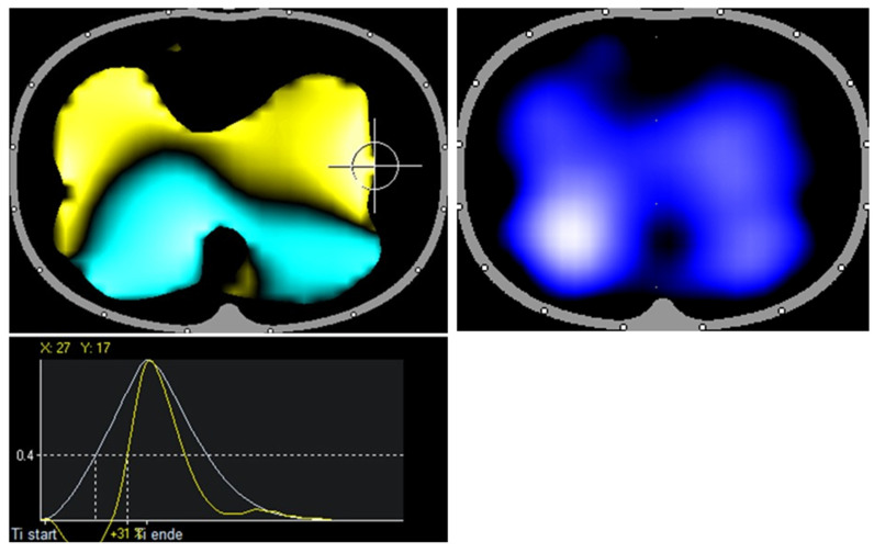
Figure 1.
Healthy volunteer: RVD image shows symmetrical ventilation to both lungs up to the marginal areas. The air first reaches the dorsal lung regions symmetrically on both sides (turquoise area) and the ventral lung regions (yellow area). Note especially the lateral symmetry and the calm distribution of the colors.

