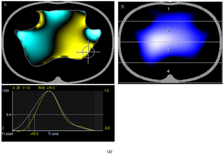
Figure 2.
(a) First presentation of the patient: Tidal image shows reduced ventilation on both ventral sides and dorsal left. The enhanced color map shows a visually recognizable irregular shape of the ventilation contour similar to thorn apple forms. Recognizable is a pronounced regional ventilation delay (left dorsal; +15 %). (b) First presentation of the patient: Largely consistent distribution of ventilation and pulsatile blood flow, except for a small right ventral right region, where pulsation but no ventilation is observed.


