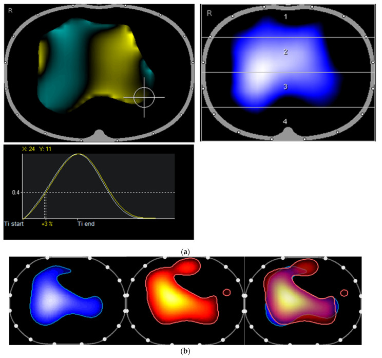
Figure 3.
(a) The second presentation of the patient: The ventilated area is at this time slightly increased with continued low ventilation (left ventral in the tidal image). The inspiratory course is more homogeneous, with only a small extent of the regional ventilation delay (left dorsal; +3%) and a lateroventrally located region that is ventilated earlier (turquoise). (b) Second presentation of the patient: The right ventral region is now ventilated again. The distribution of ventilation and pulsatile blood flow are highly concordant.

