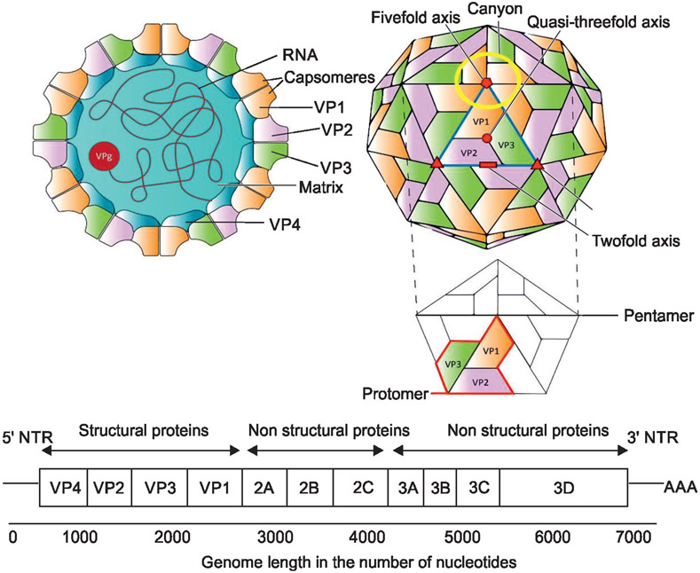
Fig. 1:
Schematic diagram showing the structure of EVs. Left-side of the schematic panel shows a cross-section with the location of the RNA, capsomeres, the matrix, and the viral proteins (VPs); Right-side of the schematic panel shows the surface with the location of structural and non-structural VPs on the surface of viral particles

