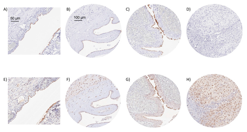
Figure 2.
IHC validation by comparison of antibodies. Using MSVA-736M, an apical membranous Upk3b positivity is seen in mesothelial cells covering an appendix (A), amnion cells of a placenta (B), and umbrella cells of the renal pelvis urothelium (C), while staining is absent in adrenal gland (D). Using clone C362, a similar membranous staining is seen in mesothelial cells of the appendix (E), amnion cells (F), and urothelial umbrella cells (G), despite of a higher level of background staining. Clone C362 also results in a significant nuclear staining of adrenocortical cells (H) which is not seen by MSVA-736M.

